Technology
Technology
Technology

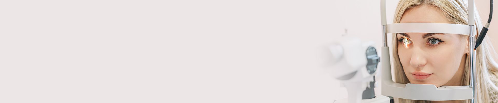
The latest technology in order to provide exceptional eye care

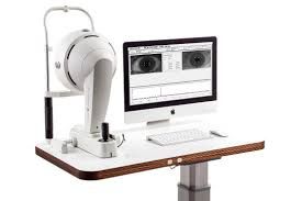
Optomap
The Optomap ultra-widefield retinal image is a unique technology that captures more than 80% of your retina in one panoramic image while traditional imaging methods typically only show 15% of your retina at one time.
The benefits of having an Optomap image taken are:
Optomap facilitates early protection from vision impairment or blindness
Early detection of life-threatening diseases like cancer, stroke, and cardiovascular diseases
The unique Optomap ultra-widefield view helps your eye doctor detect early signs of retinal disease more effectively and efficiently than with traditional eye exams. Early detection means successful treatments can be administered and reduces the risk to your sight and health.
We then have these images networked into all our exam rooms so that we can show the photos to patients to better educate them about their eye health or eye disease. Our doctors recommend the Optomap images be captured each and every year to aid in early detection of systemic and eye diseases.
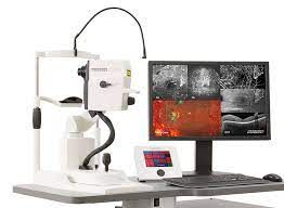
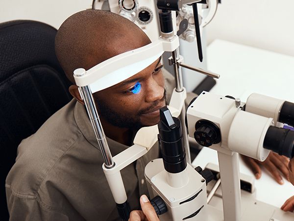
SPECTRALIS OCT
Optical coherence tomography (OCT) is an imaging method that uses light to scan the retina. It can be performed on undilated pupils and is painless. It provides detailed, real-time information about the structure of the living eye. Using light to scan the retina and optic disc, this pioneering technology brings new clinical tools for the diagnosis and management of retinal disease and glaucoma.
The Spectralis OCT allows our doctors to visualize retinal structures, evaluate the retinal thickness in 3-D and to view high-definition cross-sectional images that reveal subtle details of retinal diseases. The Spectralis OCT provides the most detailed scan patterns and layer maps available for identifying retinal and glaucoma disease characteristics and monitoring disease progression. It enables our doctors to project future vision loss and make timely treatment decisions. Our doctors use OCT technology to diagnose and monitor diseases such as glaucoma, macular degeneration, macular holes, diabetic retinopathy, other macular defects, and more.
We are proud to be the first and only eye care practice in the greater Wichita Falls area to have this latest and most up-to-date OCT technology. We feel it is very important to keep up-to-date by providing the latest in cutting-edge technology to better serve your eye care needs.
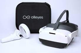
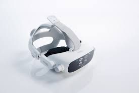
Visual Field Analysis
Visual fields are used to check a patients’ peripheral (or side) vision. Visual field loss may occur due to diseases or disorders of the eye, optic nerve, or brain. We measure this by using a virtual reality headset complete with a multi-lingual virtual assistant to perform a full visual field analysis where points of light are flashed onto a white screen and the patient is asked to press a button if he or she sees it. The technology computes the results and creates a map of the patient’s visual field.
Our doctors use visual field exams to diagnose and monitor glaucoma, detection of central or peripheral retinal disease, eyelid conditions such as ptosis or drooping, optic nerve disease, and diseases affecting the visual pathways within the brain.
Oculus Topographer
Corneal topography is a special photography technique that maps the surface of the clear front part of the eye called the cornea. It works much like a 3D map of the world, that helps identify mountains and valleys. It maps out the surface of the eye.
Our doctors use corneal topography to diagnose various conditions such as keratoconus, dry eye, and other corneal irregularites.

Specular Microscope
Konan specular microscope provides one of the most sensitive indicators of the health of the cornea by providing cellular level views of the endothelium. The specular microscope assesses cell density, cell size variation, cell shape variation, and corneal thickness.

LipiView
LipiView is the only instrument available that measures lipid layer thickness, evaluates the blink dynamics and images meibomian gland structure for your doctor to evaluate the level at which your glands are or are not functioning.
The LipiView accurately measures the sub-micron thickness of the lipid layer. LipiView analyzes over one billion data points, isolating the lipid layer of the tear film to generate an accurate repeatable measurement.

LipiFlow
While there are multiple choices available for treating MGD, LipiFlow is the only FDA-cleared device for removing gland blockages and restoring gland function. Through advances in the application of Vectored Thermal Pulsation (VTP) technology, the LipiFlow treatment utilizes a patented algorithm of heat applied to the inner eyelids and massage to remove the obstructions in your meibomian glands.
AdaptDx by Maculogix
The AdaptDx®, created by MacuLogix® measures the number of minutes it takes for the eye to adapt from bright light to darkness—the Rod Intercept™ (RI). With the AdaptDx multi-lingual virtual reality headset, you get a clear, objective measurement of retinal function with 90% sensitivity and 90% specificity for the presence of AMD. Diagnosing our patients that have AMD (age-related macular degeneration) early allows our doctors a course of action to delay or prevent blindness.


(940) 691-5653
Our fax number is able to receive text messages too!
- Monday10:00 AM - 5:30 PM
- Tuesday8:00 AM - 5:30 PM
- Wednesday8:00 AM - 5:30 PM
- Thursday8:00 AM - 5:30 PM
- Friday8:00 AM - 4:00 PM
- SaturdayClosed
- SundayClosed
(940) 569-4648
- Monday1:00 PM - 5:30 PM
- Tuesday8:00 AM - 5:30 PM
- Wednesday8:00 AM - 5:30 PM
- Thursday8:00 AM - 5:30 PM
- FridayClosed
- SaturdayClosed
- SundayClosed
Powered by: ![]()